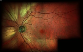

MYELINATED RETINAL NERVE FIBER LAYER PATCH
Histopathological analysis demonstrates that myelinated fibers are not confined to a patch or fascicle, and single myelinated fibers can be found in between fascicles of unmyelinated fibers. Although the exact causes remain unknown, MRNF occur when the myelination process extends past the lamina cribrosa and is visible on fundus examination. Normally, the myelination process stops at this level some postulated explanations for this observation are the presence of the structure of the lamina cribrosa, the leakage of plasma proteins from the choroidal circulation inducing the differentiation of oligodendrocytes, and factors from type 1 astrocytes blocking oligodendrocyte migration. Myelination along the visual pathway is first seen in the eighth month of gestation, and typically reaches the posterior globe around the time of birth with virtually all fibers reaching complete myelination by age 7 months. Retinal ganglion cell myelination proceeds from the lateral geniculate nucleus anteriorly toward the globe. PathophysiologyĪxonal myelination in the human central nervous system is a complex, orderly process carried out by oligodendrocyte progenitor cells, which migrate under the influence of neuro-hormonal signals to generate oligodendrocytes that produce myelin. In the other eye, I found, without much surprise, in the same place, the ring around the disc with a width of 2–2, 5 mm, regressing towards the outside”. Macula was normal and near the optic disc, though more deeply situated, were thick, opaque, chalk-white spots, which spread around the disc in the shape of a star, so that when I wanted to draw the line between the disc and macula on each side of the two had the same divergence. The German pathologist Rudolf Virchow was the first to describe myelinated retinal nerve fibers, writing in 1856 that the “retina was white, very thick and wrinkled. ICD-10 H35.89 Defect, defective retinal nerve bundle fibers ICD-9 377.49 Other disorders of optic nerve Disappearance of MRNF have also been reported after surgery and insults to the optic nerve. MRNF are typically present at birth and are static lesions, but a few cases of acquired and progressive lesions in both childhood and adulthood have been described. Though rare, familial cases of MRNF have been reported both in isolation and in combination with ocular and systemic syndromes. Most patients with MRNF are asymptomatic however, some patients have associated ocular findings including axial myopia, amblyopia, and strabismus. MRNF are present in 0.57 to 1% of the population and can occur bilaterally in approximately 7% of affected patients. Clinically, they appear to be gray-white well-demarcated patches with frayed borders on the anterior surface of the neurosensory retina. Myelinated retinal nerve fiber layers (MRNF) are retinal nerve fibers anterior to the lamina cribrosa that, unlike normal retinal nerve fibers, have a myelin sheath. 6.4 Conditions associated with loss of myelination of the RNFL.6.3 Conditions associated with acquired and progressive myelination of the RNFL.6.2 Systemic associations with myelination of the RNFL.6.1 Ocular associations with myelination of the RNFL.6 Conditions associated with myelination of the RNFL.1.1 International Classification of Disease.


 0 kommentar(er)
